Anatomy Of Chest Bones - Https Www Thoracic Theclinics Com Article S1547 4127 10 00125 8 Pdf : It describes the theatre of events.. This framework consists of many individual bones and cartilages. Language and terminology for the study of the anatomy of the thorax. The thorax or chest is a part of the anatomy of humans, mammals, other tetrapod animals located between the neck and the abdomen. Clavicle bone is a narrow rod like s shaped bone extending between sternum and acromion process of scapula bone. There also are bands of fibrous connective tissue—the ligaments and the tendons—in intimate relationship with the parts of the skeleton.
Ground substance and collagen fibers create a matrix that contains. All of the anatomical and important histological facts about the bones, together with the clinical relations, are going to be desrcibed in this article. The former is a type of connective tissue made up of cells suspended in a matrix: The thorax or chest is a part of the anatomy of humans, mammals, other tetrapod animals located between the neck and the abdomen. Learn about each muscle, their locations & functional anatomy.
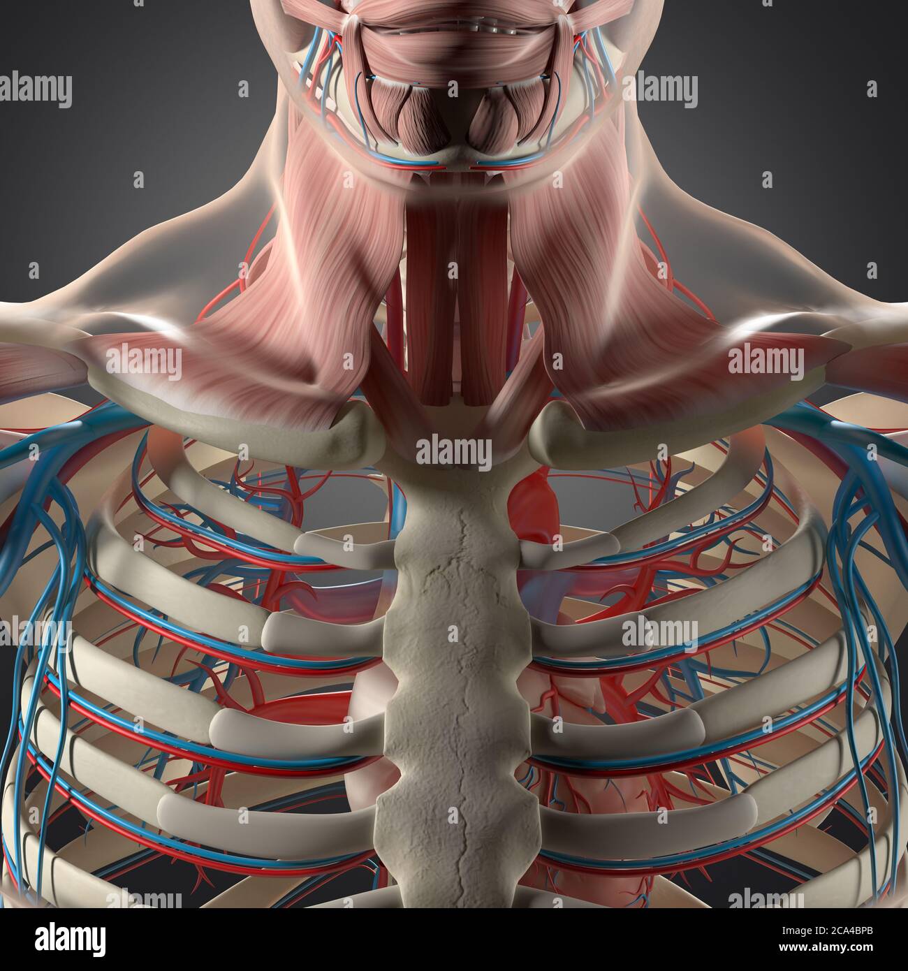
Human Anatomy Illustration Chest Rib Cage Vascular System 3d Illustration Stock Photo Alamy from c8.alamy.com Click to view large image. This framework consists of many individual bones and cartilages. There also are bands of fibrous connective tissue—the ligaments and the tendons—in intimate relationship with the parts of the skeleton. All of the anatomical and important histological facts about the bones, together with the clinical relations, are going to be desrcibed in this article. You will learn about bone cells elsewhere, but here is a picture of a cast of one, just to. Bone basics and bone anatomy. Right upper anatomy is to physiology as geography is to history: Long bones are categorised by their tubular shaft (diaphysis) with a rounded end (epiphysis) on each end.
Identify the following structures on the lateral chest radiograph:
Learn skull anatomy with skull bones quizzes and diagram labeling exercises. Hand | definition, anatomy, bones, diagram, & facts. This is an updated version of the 2007 article. Anatomists talk about both bone and bones. It originates at your clavicle, ribs, and sternum, and inserts into the upper portion of your humerus (upper arm bone from elbow to shoulder.) In this review we present the normal axial and coronal anatomy of the temporal bone by scrolling through the images. There also are bands of fibrous connective tissue—the ligaments and the tendons—in intimate relationship with the parts of the skeleton. These bones form by the thickening of a. 12 photos of the anatomy bones chest. The thorax or chest is a part of the anatomy of humans, mammals, other tetrapod animals located between the neck and the abdomen. Long bones function to support the weight of the body and facilitate movement. Click to view large image. The bones have a the scapula, or shoulder blade, is an approximately triangular shaped bone.
Clavicle bone is a narrow rod like s shaped bone extending between sternum and acromion process of scapula bone. This is an updated version of the 2007 article. Ground substance and collagen fibers create a matrix that contains. Long bones are categorised by their tubular shaft (diaphysis) with a rounded end (epiphysis) on each end. Sesamoid bones are generally small, flat and have an apex at one end.
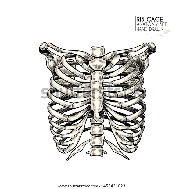
Manubrium Hd Stock Images Shutterstock from image.shutterstock.com It is made up of the wrist joint, the carpal bones, the metacarpal bones, and the phalanges. It originates at your clavicle, ribs, and sternum, and inserts into the upper portion of your humerus (upper arm bone from elbow to shoulder.) 12 photos of the anatomy bones chest. This framework consists of many individual bones and cartilages. Sesamoid bones are generally small, flat and have an apex at one end. It describes the theatre of events. Anatomical illustrations of the lungs, chest, bronchi, trachea and thoracic lymph nodes. This webpage presents the anatomical structures found on wrist mri.
Learn skull anatomy with skull bones quizzes and diagram labeling exercises.
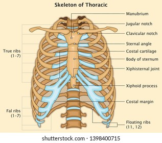
The manubrium, sternal body, and xiphoid process. Long bones are categorised by their tubular shaft (diaphysis) with a rounded end (epiphysis) on each end. The bones have a the scapula, or shoulder blade, is an approximately triangular shaped bone. Right upper anatomy is to physiology as geography is to history: They are always longer than they are wide the vertebrae are irregular bones. Where is the sternum found. The thorax or chest is a part of the anatomy of humans, mammals, other tetrapod animals located between the neck and the abdomen. They are collectively known as the tarsus. Long bones are mostly located in the appendicular skeleton and include bones in the lower limbs (the tibia, fibula, femur, metatarsals, and phalanges) and bones in the upper limbs (the humerus, radius, ulna, metacarpals. Anatomy of the brain | model. Ground substance and collagen fibers create a matrix that contains. This framework consists of many individual bones and cartilages. Talus calcaneus navicular cuboid lateral cuneiform intermediate cuneiform medial cune.
Long bones function to support the weight of the body and facilitate movement. Compare the nuclear medicine scans to anatomical diagrams. Learn about each muscle, their locations & functional anatomy. Where is the sternum found. Vascular anomalies of aorta, pulmonary and systemic vessels.
Hand Drawn Anatomy Set Vector Human Stock Vector Royalty Free 1453431023 from image.shutterstock.com The medial anterior chest is defined by the sternum, which consists of 3 flat polygonal bones: Learn skull anatomy with skull bones quizzes and diagram labeling exercises. They are always longer than they are wide the vertebrae are irregular bones. Bone comprises the structure of the skeletal system and provides lever arms for locomotion. Sesamoid bones are generally small, flat and have an apex at one end. A bone is a somatic structure that is comprised of calcified connective tissue. This anatomical midline can be useful in assessing for symmetry in breast augmentation or in performing a median sternotomy. It describes the theatre of events.
Click to view large image.
Where is the sternum found. This framework consists of many individual bones and cartilages. Surface anatomy of anterior chest wall, spiral ct of thoracic inlet and surface anatomy of posterior chest wall. Breast bone anatomy human breast bone anatomy bone anatomy sternum | chest bone : Learn about each muscle, their locations & functional anatomy. It originates at your clavicle, ribs, and sternum, and inserts into the upper portion of your humerus (upper arm bone from elbow to shoulder.) There also are bands of fibrous connective tissue—the ligaments and the tendons—in intimate relationship with the parts of the skeleton. This anatomical midline can be useful in assessing for symmetry in breast augmentation or in performing a median sternotomy. O bones—spine, ribs, clavicles, scapulae, humeri. Identify the following structures on the lateral chest radiograph: Click to view large image. Ground substance and collagen fibers create a matrix that contains. Anatomists talk about both bone and bones.
This framework consists of many individual bones and cartilages anatomy of chest. Bone of chest and their parts.


0 Komentar